![]()
Rev. Ciencias Veterinarias, Vol. 37, N° 2, [1-10], E-ISSN: 2215-4507, julio-diciembre, 2019
DOI: https://doi.org/10.15359/rcv.37-2.1
URL: http://www.revistas.una.ac.cr/index.php/veterinaria/index
Ultrasonographic measurement of the equine fetal vitreous body length for predicting days to parturition in Pura Raza Española horses
Medición ultrasonográfica del largo del cuerpo vítreo fetal equino para predecir los días para el parto en caballos Pura Raza Española
Medição ultrassonográfica do comprimento do corpo vítreo fetal equino para prever os dias para o parto em cavalos Pura Raça Espanhola
Patricio Razquin-Echeverriarza1  , Patrick M. McCue2, Paula Cappella-Flores3, Bernardo Vargas-Leitón1, Sandra Estrada-König1
1 School of Veterinary Medicine, Universidad Nacional, Heredia, Costa Rica. Emails: patorazquin@gmail.com, bernardo.vargas.leiton@una.cr , sandrae@una.cr
2 Department of Clinical Sciences, Colorado State University, Fort Collins, Colorado, USA. Email: patrick.mccue@colostate.edu
3 Private practitioner, Heredia, Costa Rica. Email: paula.cappella@gmail.com
: Patricio Razquin, patorazquin@gmail.com
Received: January 14, 2019. Corrected: June 4, 2019. Accepted: June 8, 2019.
Abstract: Ultrasonographic measurement of the fetal vitreous body length to predict parturition date in horses has shown substantial differences between breeds. PRE (Pura Raza Española or Purebred Spanish Horse) is an important breed in the equine industry of Costa Rica. No data for prediction of parturition exists for PRE using fetal ocular measurements. Between-observer agreement has never been evaluated for fetal ocular measurements on horses. A total of 86 ocular diameters were measured by a veterinarian in twelve PRE mares from day 240 of gestation until parturition. Forty measurements were repeated by a senior veterinary student to determine between-observer agreement. Transrectal ultrasonography was performed in each occasion, and a mean was calculated of the three measurements obtained. Two nonlinear regression equations were derived using days before parturition and age of gestation as dependent variables and vitreous body length as the independent variable. The model obtained for days before parturition was y = 1123.8 -55.5*x+0.689*x2, where “y” represents days before parturition and “x” represents fetal ocular diameter (r2= 0.79; P<0.001); and y = -710.6+51.8*x -0.644*x2, where “y” represents age of gestation and “x” represents ocular diameter (r2= 0.75; P<0.001). Pearson correlation coefficient and paired t-test were performed to assess the between-observer agreement. No significant differences (p=0.86) were detected between-observers, indicating high reproducibility. This study concluded that ocular diameter measurement can be reproduced with high precision by different veterinarians, and either model using days before parturition or age of gestation can be used to predict parturition in PRE mares when breeding dates are unknown.
Keywords: Pura Raza Española, Mares, Ocular, Diameter, Parturition, Ultrasonography
Resumen: La medición ultrasonográfica del largo del cuerpo vítreo fetal como forma de predicción de la fecha del parto ha mostrado diferencias substanciales entre especies. Los pura raza española (PRE) son una raza de importancia económica en la industria ecuestre de Costa Rica. No existen datos para la predicción del parto utilizando esta medición para la raza PRE. El acuerdo entre observadores no ha sido evaluado para las mediciones oculares en fetos equinos. Un total de 86 diámetros oculares fueron medidos por un veterinario en 12 yeguas PRE desde el día 240 de gestación hasta el parto. Cuarenta mediciones fueron repetidas por un estudiante de veterinaria de último año para determinar el acuerdo entre observadores. En cada ocasión, se realizó ultrasonografía transrectal, en las cuales se obtuvieron tres mediciones con las cuales se calculó un promedio. Dos regresiones no lineares fueron derivadas utilizando días para el parto y edad gestacional como variables dependientes y largo del cuerpo vítreo como variable independiente. El modelo obtenido para días para el parto fue y = 1123.8 -55.5*x+0.689*x2 , donde “y” representa los días para el parto y “x” representa la medida del diámetro ocular (r2= 0.79; P<0.001); y y = -710.6+51.8*x -0.644*x2, donde “y” representa la edad gestacional y “x” representa el diámetro ocular (r2= 0.75; P<0.001). Se realizaron las pruebas de coeficiente de correlación de Pearson y T test pareada para evaluar el acuerdo entre observadores. No se detectaron diferencias significativas (p=0.86) entre los observadores, indicando una alta reproducibilidad de la medida. Este estudio concluyó que la medición del diámetro ocular puede ser reproducida con alta precisión por diferentes veterinarios, y ambos modelos utilizando días para el parto o edad gestacional pueden ser utilizados para predecir el parto en yeguas PRE cuando la fecha del servicio es desconocida.
Palabras clave: Pura raza española, yeguas, ocular, diámetro, parto, ultrasonografía
Resumo: A medida ultrassonográfica do comprimento do corpo vítreo fetal como forma de prever a data do parto tem mostrado diferenças substanciais entre as espécies. A Pura Raça Espanhola (PRE) é uma raça de importância econômica na indústria equestre da Costa Rica. Não há dados para a previsão do parto usando esta medida para a raça PRE. A concordância entre observadores não foi avaliada para medidas oculares em fetos equinos. Um total de 86 diâmetros oculares foram medidos por um veterinário em 12 éguas PRE desde o dia 240 da gestação até o parto. Quarenta medições foram repetidas por um estudante de último ano do curso de medicina veterinária, para determinar a concordância entre os observadores. Em cada ocasião, foi realizada ultrassonografia transretal, na qual foram obtidas três medidas com as quais foi calculada uma média. Duas regressões não lineares foram obtidas utilizando-se os dias para o parto e a idade gestacional como variáveis dependentes e o comprimento do corpo vítreo como variável independente. O modelo obtido para os dias para o parto foi y = 1123,8 -55,5 * x + 0,689 * x2, onde “y” representa os dias para o parto e “x” representa a medida do diâmetro ocular (r2 = 0,79; P <0,001) ; e y = -710,6 + 51,8 * x -0,644 * x2, onde “y” representa a idade gestacional e “x” representa o diâmetro ocular (r2 = 0,75; P <0,001). O coeficiente de correlação de Pearson e o teste “t” pareado foram realizados para avaliar a concordância entre os observadores. Não foram detectadas diferenças significativas (p = 0,86) entre os observadores, indicando alta reprodutibilidade da medida. Este estudo concluiu que a medição do diâmetro ocular pode ser reproduzida com alta precisão por diferentes médicos veterinários, e ambos os modelos que utilizam dias para o parto ou idade gestacional podem ser usados para prever o parto em éguas PRE, quando a data da inseminação ou monta natural é desconhecida.
Palavras-chave: Pura raça espanhola, éguas, ocular, diâmetro, parto, ultrassonografia.
Introduction
The use of ultrasonographic ocular measures to determine age of gestation in mares of different breeds has been assessed by different authors, with the purpose of obtaining an approximate date of parturition as precise as possible to implement management measures and improve foal survival rate. Different fetal eye measurements such as the sum of width and length (Renaudin et al. 2000; Bucca et al. 2005; Turner et al. 2006; Hendriks et al. 2009), approximate eye volume (Renaudin et al. 2000; Hendriks et al. 2009), and vitreous body length (Kahn & Leidl 1987; Turner et al. 2006; Hartwig et al. 2012) have been studied, the latter being the best single predictor for age of gestation (Turner et al. 2006).
Differences in growth patterns were described in various studies, evidencing either a linear growth pattern in Crioulo horses (Hartwig et al. 2012), light horses (Kahn & Leidl 1987), and standardbreds, (Lanci et al. 2018) or a curvilinear pattern in Dutch Warmbloods (Hendriks et al. 2009), light horses, ponies (Turner et al. 2006), and Thoroughbreds (Murase et al. 2014). Although different growth patterns may be due to the methodology implemented in each study, an evident variation in size and development pattern of the fetal ocular orbit has been pointed out, even in breeds of similar size (Kahn & Leidl 1987; Renaudin et al. 2000; Turner et al. 2006; Hendriks et al. 2009, Hartwig et al. 2012). Since there is no data for every breed, extrapolation from horses of similar sizes has been a common practice in field veterinarians, leading to not very accurate predictions of parturition.
Pura Raza Española (PRE), an important breed in Costa Rica, corresponds to a significant proportion of the equine industry in the country, with 3000 animals registered as of 2017 and an exponential growth in the last few years. Although most of the breeding farms have competent reproductive management and records, there are still situations where the age of gestation in a mare is unknown, creating an unnecessary risk at parturition, both for foal and mare.
Reproducibility is a measure of reliability, which has the purpose of assessing the between-observer agreement (Watson & Petrie 2010). Even though the vitreous body length measure is used constantly by field veterinarians, to our knowledge, the reproducibility of the measure has not been assessed.
The objective of this study was to develop reference ranges for prediction of parturition in PRE mares using vitreous body length measured by transrectal ultrasonography of the fetus from 270 days of gestation until parturition, and to assess the between-observer agreement of this method.
Materials and Methods
Twelve PRE mares between five and 14 years old, with gestations of 240 to 339 days, were used for the study. Mares weighed between 450 kg and 600 kg, had body scores greater than five (Henneke et al. 1983), and were kept in three breeding farms in the provinces of Heredia and San José, Costa Rica. These three breeding farms had similar nutrition and reproductive management, and every mare had had an annual breeding soundness examination before entering the reproduction program. Mares were kept in paddocks and had pasture and water ad libitum in addition to grain. Ultrasound examinations were performed daily when signs of estrus were evident, and ovulation was defined as the absence of a dominant follicle that was present on the previous examination. The day of ovulation was defined as day zero, and ultrasonography was performed at days 14 and 35 to check for pregnancy. Mares were mated either by artificial insemination or natural breeding.
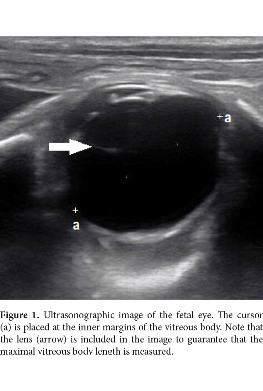
A total of 86 fetal ocular diameters were measured in the 12 mares, from day 240 of pregnancy to parturition (mean gestational length 340 days; SD= 8.86; range= 323 to 360), with a period between measurements of two to four weeks depending on availability of the mares. Three measures of ocular diameter were made by the same operator and an average was calculated. A transrectal ultrasonography examination was conducted in every mare using stocks as means of restraint. Ultrasounds were performed using a portable ultrasound unit equipped with 7.5 mHz linear array transducer (Mindray M5, Shenzhen Mindray Bio-Medical Electronics Co., Ltd.). To localize the fetal orbit, the transducer was moved from left to right over the uterus, starting at the level of the cervix until the orbit was found. Then the transducer was moved over the orbit to get an image where the longest axis was measured, using the inner margins of the vitreous body and lens as landmarks (Figure 1). Three images were saved every time, and measures were taken of all three.
For the analysis of the between-observer agreement, 40 measures of the fetal ocular diameter were made by an experienced veterinarian and repeated by a senior veterinary student. Measures were blinded so each observer did not have access to the other measurements, and both operators measured each ocular diameter three times.
Every parturition date was recorded, and normal healthy foals were obtained from all 12 pregnancies, including seven colts (mean gestation length at day 335) and five fillies (mean gestation length at day 343).
Statistical Analysis
Descriptive statistics (mean, standard deviation, and 95% confidence intervals) were obtained for quantitative data. Pearson correlation coefficients were calculated for the three measurements of ocular diameter, both between and within observers. The difference in average measurements taken by the two observers in the same mares was also analyzed using a paired sample t-test. Once the parturition dates were known, days before parturition at the time of measurements were calculated for each mare. Two different nonlinear regression models were derived using days before parturition and age of gestation as dependent variables and vitreous body length as the independent variable. Days to parturition were calculated from data obtained from the model using age of gestation as the dependent variable (considering 340 days as average gestation) and a comparison was made with the model using days before parturition as the dependent variable. Statistical assumptions for the regression model (normality, homoscedasticity, and independence of errors) were graphically assessed. All statistical analyses were made using SAS 9.6 software (SAS Institute Inc., Cary, NC).
Results and Discussion
Between-Observer Agreement
Of the 86 ocular diameters measured, 90% successfully obtained agreed with the data from other studies (Turner et al. 2006). Only in two occasions (5%) was the inexperienced veterinary student unable to measure the ocular diameter, while the veterinarian could; and in two examinations (5%) neither one was able to measure the ocular diameter, probably because of a low position of the head of the fetus. Minimum variation between measures of the same operator was 0.1 mm while maximum variation was 3.1 mm (mean 0.7 mm). Maximum variations between measures of the same ocular diameter were observed in situations where the inner margins of the vitreous body were not completely defined, so the operator had to guess the borders in order to make a measurement. Although this was not a common problem, it resulted in the highest variation. Turner et al. 2006 demonstrated that the predictive value of three versus two measurements was slightly increased and probably reduces the error when problems detecting scleral limits are encountered. In every examination where an ocular diameter was detected, all the three measurements were made with no complications. Pearson correlation coefficient was highly significant (0.96; p<0.0001) for the three measurements within each observer.
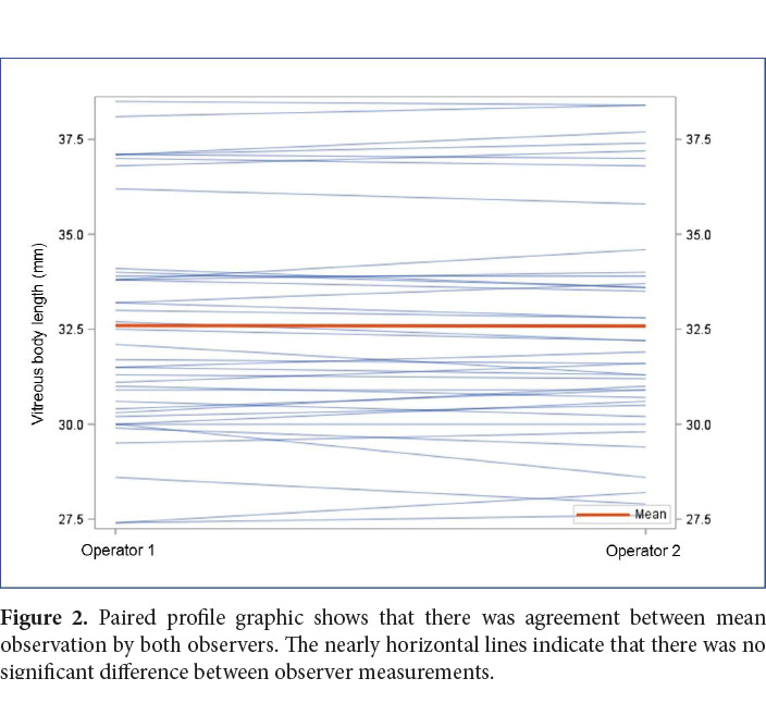
For reproducibility assessment, minimum and maximum variations between observers were 0.4 mm and 2.5 mm, respectively (mean 1 mm). Pearson coefficient was significant (0.95; p<0.0001) for ocular diameter measures from both operators. To account for agreement between both observers, t-test was performed and no significant difference was detected (p=0.86; Mean= 0.0128) (Figure 2).
To our knowledge, between-observer agreement has not been evaluated before for measurements of vitreous body length. Due to the small difference between observers detected in our study, where a student was used to compare measurements with an experienced veterinarian, we can conclude that this measure can be most likely repeated by field veterinarians, with enough precision for results to be useful. Other factors, such as difference in ultrasound machine, were not included in our study and could be a source or variation, but further research is needed to account for that.
Regression Model
Two different approaches have been used for the prediction of parturition in horses using ocular measurements. Turner et al. 2006 used days before parturition to predict parturition date in pony mares, while several other authors have used age of gestation (Kahn & Leidl 1987; Hendriks et al. 2009; Hartwig et al. 2012). The days before parturition approach takes into consideration the natural variation in gestation length, while age of gestation is the common predictor of foaling when ovulation date is known; therefore, clinicians are more familiar with the last parameter.
Figure 3 represents a nonlinear (quadratic) regression model to determine the age of gestation of PRE mares using days before parturition as dependent variable. The predictive equation derived was y= 1123.8 -55.5*x+0.6898*x2, where “y” represents days before parturition and “x” represents vitreous body length in millimeters. Overall adjustment of this model was highly significant (P<0.001, r2= 0.79 RMSE= 15.3 days).
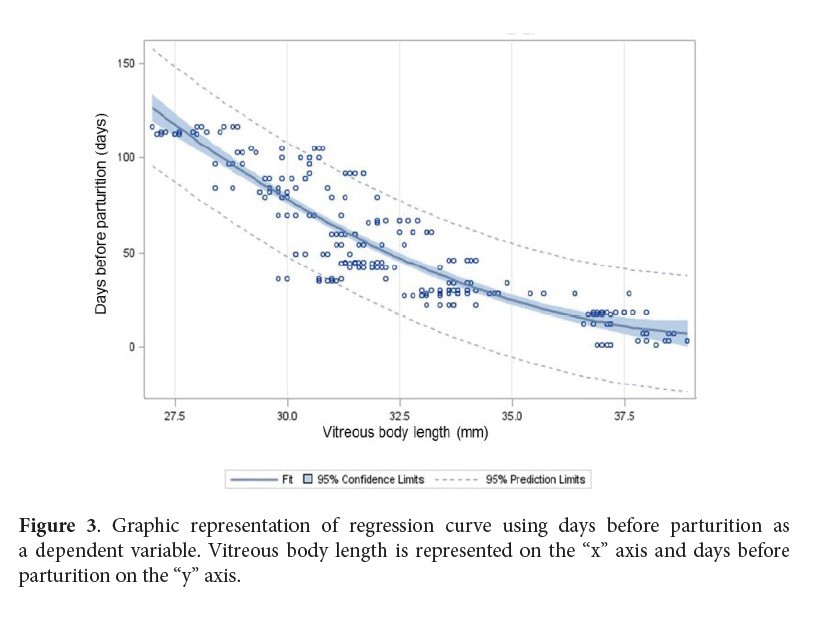
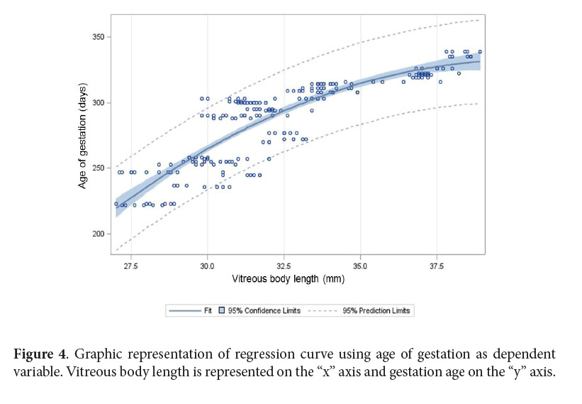
Figure 4 represents the nonlinear (quadratic) regression model using age of gestation as dependent variable. The predictive equation derived was y= -710.6+51.8*x-0.644*x2, where “y” represents age of gestation and “x” represents vitreous body length in millimeters. The significance of this model was high (P<0.001, r2= 0.76 RMSE= 15.7 days). Graphical assessment indicated no significant deviation from statistical assumptions in both models.
Hendriks et al. 2009 described that the fetal ocular diameter increased linearly until approximately day 240 of gestation in 32 Dutch warmbloods. Thereafter, growth slowed down, and a plateau was reached around day 300. Turner et al. 2006 also described a linear growth pattern in ponies until day 280, where a curvilinear pattern was presented. This is in contrast with Hartwig et al. 2012, who described a linear growth pattern in crioulo mares until parturition. Kahn & Leidl 1987 also described a linear growth pattern, but the number of measures contributing to their model after day 240 of gestation was small (six out of 96).
In our models, a curvilinear growth pattern is observed from day 240 to parturition, which coincides with the data from other equine breeds, but a plateau was not evident in the last month of gestation, when there was a continuous growth until parturition. This result could be explained by a variation in the growth pattern in PRE horses; however, other factors not taken into account in our study such as parity, mare size, and different lineages may be influencing the results. No measurements were conducted in our study before day 220. Earlier measurements may have demonstrated a linear growth pattern as described by other authors (Kahn & Leidl 1987; Turner et al. 2006; Hendriks et al. 2009).
Table 1 lists prediction intervals for PRE mares. Although there is a significant correlation between days before parturition, age of gestation, and vitreous body length, a relatively wide prediction interval resulted from our model. For this reason, ocular measurements should be used as an approximation, and other indicators of parturition proximity should be used in conjunction for a more accurate prediction.
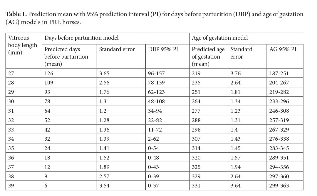
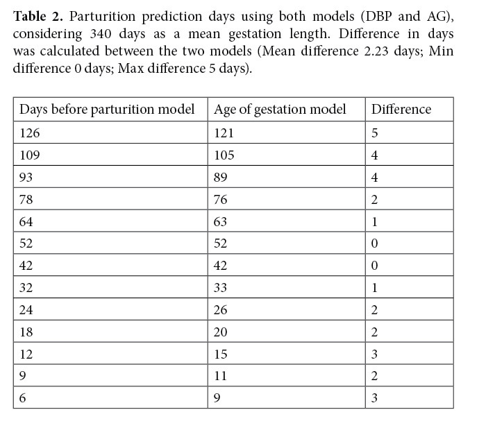
Table 2 shows the difference in days for prediction of parturition using both models. Due to the small difference, any of the two models is suitable for calculating foaling date, although some factors should be considered. Pregnancy length has been reported to vary from 320 to 362 days (Rollins, 1951), which may lead to an increase in days to parturition up to 20 or more days if age of gestation is considered. Although the days before parturition model takes into account the gestation variation, no considerable difference was obtained when comparing both models, probably because our own gestation length mean was 340 days.
The comparison with other models has demonstrated that significant differences in the growing pattern exist between different horse breeds of similar size, probably because of the eye to skull width ratio that may vary as a characteristic of breed (Turner et al. 2006; Hartwig et al. 2012) accounting for the importance of breed specific data. Other factors have been studied for a relationship with the eye growth pattern, like number of foaling (primiparous vs multiparous) (Turner et al. 2006; Hendriks et al. 2009), maternal age (Allen et al. 2002; Hendriks et al. 2009), foal gender, body condition, and size of mares (Turner et al. 2006). None of these variables were included in our study due to sample size restrictions; therefore, variations in the growth curve could exist associated with these factors.
Conclusion
In conclusion, the fetal ocular diameter is a simple, repeatable, and reproducible measure that can aid in the determination of a parturition date in PRE mares. Difference in models using days before parturition or age of gestation were not considerable in this study; consequently, either approach could be used by field veterinarians. Breed specific work is still needed to determine growth curves in other breeds.
Conflict of Interest
None.
References
Allen, W.R., Wilsher S., Stewart, F., Ousey, J. & Fowden, A. 2002. The influence of maternal size on placental, fetal and postnatal growth in the horse. II. Endocrinology of pregnancy. J Endocrinol; 172:237–46. doi:10.1677/joe.0.1720237
Bucca, S., Fogarty, U., Collins, A. & Small, V. 2005. Assessment of feto-placental well-being in the mare from mid-gestation to term: Transrectal and transabdominal ultrasonographic features. Theriogenology; 64:542–57. doi: 10.1016/j.theriogenology.2005.05.011
Hartwig, F.P., Antunez, L., Dos Santos, R.S., Lisboa, F.P., Pfeifer, L.F.M. & Nogueira, CEW. 2012. Determining the gestational age of crioulo mares based on a fetal ocular measure. J Equine Vet Sci; 33:557–60. doi: 10.1016/j.jevs.2012.08.203
Hendriks, W.K., Colenbrander, B., Van der Weijden, G.C. & Stout, T.A.E. 2009. Maternal age and parity influence ultrasonographic measurements of fetal growth in Dutch Warmblood mares. Anim Reprod Sci; 115:110–23. doi: 10.1016/j.anireprosci.2008.12.014
Henneke, D.R., Potter, G.D., Kreider, J.L. & Yeates, B.F. 1983. Relationship between condition score, physical measurements and body fat percentage in mares. Equine Vet J; 15:371–2. doi: https://doi.org/10.1111/j.2042-3306.1983.tb01826.x
Kahn, V.W. & Leidl, W. 1987. Die ultraschall-biometrie von pferdefeten in utero und die sonographische darstellung ihrer organe. Dtsch Tierarztl Wschr; 94:497–540
Lanci, A., Castagnetti, C., Ranciati, S., Sergio, C. & Mariella, J. 2018. A regression model including fetal orbit measurement to predict parturition in Standardbred mares with normal pregnancy. Theriogenology; 126:153–158. https://doi.org/10.1016/j.theriogenology.2018.12.020
Murase, H., Endo, Y., Tsuchiya, T., Kotoyori, Y., Shikichi, M. & Ito, K. 2014. Ultrasonographic Evaluation of Equine Fetal Growth Throughout Gestation in Normal Mares Using a Convex Transducer. J Vet Med Sci; 76:947–53. doi:10.1292/jvms.13-0259
Renaudin, C.D., Gillis, C.L., Tarantal, A.F. & Coleman, D.A. 2000. Evaluation of equine fetal growth from day 100 of gestation to parturition by ultrasonography. J Reprod Fertil; 56:651–60.
Rollins, C.E. & C. H.W. 1951. Environmental Sources of Variation in the Gestation Length of the Horse. J Anim Sci; 10:789–796. doi:https://doi.org/10.2527/jas1951.104789x
Turner, R.M., McDonnell, S.M., Feit, E.M., Grogan, E.H. & Foglia, R. 2006. Real-time ultrasound measure of the fetal eye (vitreous body) for prediction of parturition date in small ponies. Theriogenology; 66. doi: 10.1016/j.theriogenology.2005.11.019
Watson, P.F. & Petrie, A. 2010. Method agreement analysis: A review of correct methodology. Theriogenology; 73:1167–79. doi: 10.1016/j.theriogenology.2010.01.003
Artículo por Revista Ciencias Veterinarias se distribuye bajo una Creative Commons Reconocimiento-NoComercial-SinObraDerivada 3.0 Costa Rica License.
Basada en una obra en http://www.revistas.una.ac.cr/index.php/veterinaria/index.
Permisos que vayan más allá de lo cubierto por esta licencia pueden encontrarse en ciencias.veterinarias.cr@una.cr.