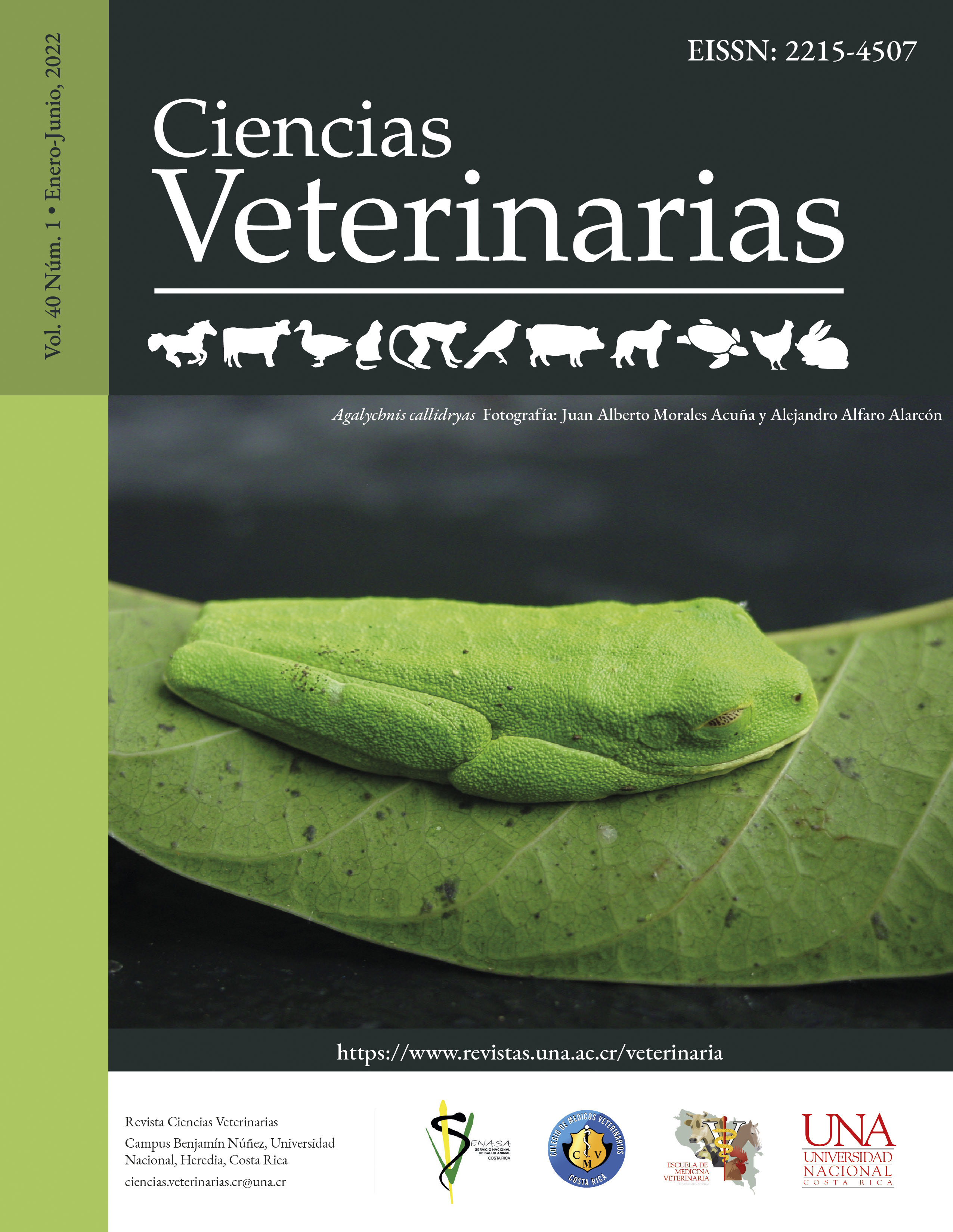
Anatomical and radiographic study on the appendicular skeleton of the Tamandua mexicana
DOI:
https://doi.org/10.15359/rcv.40-1.1Keywords:
Osteology, Xenarthra, appendicular skeleton, double scapular spine, supratrochlear foramenAbstract
Tamandua mexicana species has an important role in the natural ecosystem as a pest controller, feeding on insects such as termites. One of the main anatomical adaptations that this species has undergone has been to its thoracic extremities. Having detailed knowledge regarding the osteology of the thoracic limbs of T. mexicana provides a strong base for its application in clinical-surgical practice. In addition to collaborating with the greater understanding of animal physiology and behavior. Because there was a lack of description about the appendicular skeleton anatomy of this species, the objective of this investigation was to describe the osteology and the radiographic anatomy of the appendicular skeleton of the T. mexicana. The bones used belonging to the appendicular skeleton of two specimens of T. mexicana were properly cleaned using standard boiling and maceration techniques. The morphometry of the bones was performed using a measuring tape, pachymeter, and radiographies. With this study, it was possible to identify and describe the anatomical peculiarities such as the presence of the double scapular spine that shapes the caudolateral fossa, and at the end of the humerus, the supratrochlear foramen, in addition to a markedly prominent medial epicondyle. In addition, a difference was observed between metacarpal bones and the phalanges of the third digit compared to the other ones, as it is significantly thicker. These findings reinforced the evidence that a certain degree of anatomical specialization is a result of an adaptation of this species to its environment and diet. The knowledge provided by research like this contributes to the improvement of surgical techniques and diagnostic approach in the species.
References
Aydin Kabakci, A. D., Buyukmumcu, M., Yilmaz, M. T., Cicekcibasi, A. E., Akin, D., & Cihan, E. (2017). An Osteometric Study on Humerus. International Journal of Morphology, 35(1), 219–226. https://doi.org/10.4067/s0717-95022017000100036
Arguedas, R., López, E. C., & Ovares, L. (2019). Bone fractures in roadkill Northern Tamandua Tamandua mexicana (Mammalia: Pilosa: Myrmecophagidae) in Costa Rica. Journal of Threatened Taxa, 11(14), 14802–14807. https://doi.org/10.11609/jott.4956.11.14.14802-14807
Brown, D. D. (2011). Fruit-Eating by an Obligate Insectivore: Palm Fruit Consumption in Wild Northern Tamanduas (Tamandua mexicana) in Panamá. Edentata, 12(1), 63–65. https://doi.org/10.5537/020.012.0110
Burton, A. M., & González, G. C. (2006). Northern-most record of the collared anteater (Tamandua mexicana) from the Pacific slope of Mexico. Revista Mexicana de Mastozoología, 10(1), 67–70. https://doi.org/10.22201/ie.20074484e.2006.10.1.142
de Souza Hossotani, C. M., & Helder Silva, L. (2016). Aspectos reprodutivos do Tamanduá-mirim (Tamandua tetradactyla Linnaeus, 1758). Revista Brasileira de Reprodução Animal, 40(3), 95–98. http://www.cbra.org.br/portal/downloads/publicacoes/rbra/v40/n3/p095-98%20(RB619).pdf
Dyce, K. M., Sack, W. O., & Wensing, C. J. G. (2009). Textbook of Veterinary Anatomy (4th ed.). Saunders.
Esser, H., Brown, D. & Liefting, Y. (2010). Swimming in the Northern Tamandua (Tamandua mexicana) in Panama, Edentata, 11, 70-72. https://doi.org/10.1896/020.011.0112
Sesoko, F.N. (2012). Estudo anatômico e imagiológico do braço e da coxa em tamanduá- bandeira (Myrmecophaga tridactyla - linnaeus, 1758) para a determinação de acesso cirúrgico [Tesis de maestría, Universidad Estadual Paulista]. https://repositorio.unesp.br/bitstream/handle/11449/99858/000696048.pdf?sequence=1&isAllowed=y
Sesoko, F.N., Rahal, S.C., Bortolini, Z., De Souza, L.P., Vulcano, L.C., Barros, F.O. & Teixeira, C.R. (2015). Skeletal morphology of the forelimb of Myrmecophaga Tridactyla. Journal of Zoo and Wildlife Medicine, 46(4), 713-722. https://doi.org/10.1638/2013-0102.1
Freitas, K. (2018). Estudo das variações anátomo-radiográficas do esqueleto do bicho-preguiça-de-garganta-marrom (Bradypus variegatus, Schinz, 1825) [Tesis de Licenciatura, Universidad Federal da Paraíba]. https://repositorio.ufpb.br/jspui/handle/123456789/12608
Göker, P., Bozkır, M. G., Yücel, A. H. & Ozandac S. (2015). An Osteometric Study of proximal and distal femur Morphology. Cukurova Medicine, 40(3), 466-473. https://doi.org/10.17826/cutf.82812
Liebich, H.G., Maierl, J., & König, H.E. (2020). Veterinary Anatomy of Domestic Mammals: Textbook and Colour Atlas. Thieme.
Lozada-Gallegos, R.A., Muñoz-García, I.C., Ovando-Fuentes, D., Rendón-Franco, E., Reyes-Delgado, F., Rocha-Martínez, N., Trejos-Salas, B.M & Villanueva-García C. (2020). Radiographic anatomy of the forelimb in the northern tamandua (Tamandua mexicana). Journal of Zoo and Wildlife Medicine. 51(2),265-274. https://doi.org/10.1638/2018-0047
Marshall, S. K., Spainhower, K. B., Sinn, B. T., Diggins, T. P., & Butcher, M. T. (2021). Hind Limb Bone Proportions Reveal Unexpected Morphofunctional Diversification in Xenarthrans. Journal of Mammalian Evolution, 28(3), 599-619. https://doi.org/10.1007/s10914-021-09537-w
Matsuo, T., Morita, F., Tani, D., Nakamura, H., Higurashi, Y., Ohgi, J., Luziga, C. & Wada, N. (2019). Anatomical variation of habitat‐related changes in scapular morphology. Anatomia, Histologia, Embryologia, 48(3), 218-227. https://doi.org/10.1111/ahe.12426
McDonald, H.G., Vizcaíno, S.F. & Bargo, M.S. (2008). Skeletal anatomy and the fossil history of the Vermilingua. In: Vizcaíno, S. F. & Loughry, W. J. (Eds.),The Biology of the Xenarthra (1 ed., pp. 64-78). University Press of Florida.
Monteiro, L.R. & Abe, A.S. (1999). Functional and historical determinants of shape in the scapula of Xenarthran mammals: Evolution of a complex morphological structure. Journal of Morphology 241, 251-263. https://doi.org/10.1111/ahe.12426:10.1002/(SICI)1097-4687(199909)241:3<251::AID-JMOR7>3.0.CO;2-7
Mora, J.M. (2000). Mamíferos silvestres de Costa Rica. EUNED. https://books.google.co.cr/books?id=4IITb9RrSFcC&pg=PA53&dq=tamandua+mexicana&hl=es&sa=X&ved=2ahUKEwiQ3-6frZjqAhUxn-AKHdTVAOIQ6AEwAHoECAEQAg#v=onepage&q=tamandua%20mexicana&f=false.
Navarrete, D. & Ortega, J.D. (2011). Tamandua mexicana (Pilosa;Myrmecophagidae). Mammalian Species 43(874), 56-63. https://doi.org/10.1644/874.1
Nyakatura, J. A. (2011). The Convergent Evolution of Suspensory Posture and Locomotion in Tree Sloths. Journal of Mammalian Evolution, 19(3), 225–234. https://doi.org/10.1007/s10914-011-9174-x
Polania-Guzmán, P. V., & Vélez-García, J. F. (2019). Gross anatomical adaptations of the craniolateral forearm muscles in Tamandua mexicana (Xenarthra: Myrmecophagidae): development of accessory muscles and rete mirabile for its arterial supply. Heliyon, 5(8), e02179. https://doi.org/10.1016/j.heliyon.2019.e02179
Queiroz, R.P. (2012). Anatomia Óssea, Muscular E Do Movimento Das Regiões Glútea E Coxa Do Tamanduá Bandeira Myrmecophaga Tridactyla (Myrmecophagidae: Pilosa) [Tesis de Maestría, Universidad Federal de Uberlândia]. https://repositorio.ufu.br/bitstream/123456789/13027/1/d.pdf
Rojano, C., Miranda, L.M. & Ávila, R.C. (2014). Manual de Rehabilitación de Hormigueros de Colombia. Colombia: Geopark Colombia. [Archivo PDF]. https://www.researchgate.net/publication/287193942_Manual_de_Rehabilitacion_de_Hormigueros_de_Colombia#fullTextFileContent
Rossi, L.F. (2013). Citogenética y biología de la reproducción en Myrmecophagidae (Xenarthra) de Argentina [Tesis Doctoral, Universidad de Buenos Aires]. http://hdl.handle.net/20.500.12110/tesis_n5301_Rossi
Rossi, L.F, Calderón, R., Alonso, F.M., Lueces, J.P., Merani, M.S. (2013). Observaciones anatómicas e histológicas del sistema reproductor masculino y femenino en Tamandua tetradactyla (Myrmecophagidae: Xenarthra). InVet, 15(1), 17-28. https://core.ac.uk/download/pdf/148102746.pdf
Rowley, B. (2015). Protocols for cleaning and articulating large mammal skeleton: Simposium Student Journal of Science & Math 2 (1), 1-27. https://digitalcommons.calpoly.edu/symposium/vol2/iss1/3/
Sandoval-Gómez, V.E., Ramírez-Chaves, H.E. & Marín, D. (2012). Registros de Hormigas Y Termitas Presentes en la Dieta de Osos Hormigueros (Mammalia: Myrmecophagidae) en Tres Localidades de Colombia. Edentata, 13(11), 1-9. https://doi.org/10.5537/020.013.0104
Superina, M., Miranda, F.R. & Abba, A.M. (2010). The 2010 Anteater Red List Assessment. Edentata, 11(12), 96-114. https://doi.org/10.5537/020.011.0201
Taylor, B.K. 1978. The anatomy of the forelimb in the anteater (Tamandua) and its functional implications. Journal of Morphology, 157(3), 347-367. https://doi.org/10.1002/jmor.1051570307
Toledo, N., Bargo, M.S. & Vizcaíno, S.F. (2015). Muscular reconstruction and functional morphology of the hindlimb of santacrucian (Early Miocene) sloths (Xenarthra, Folivora) of Patagonia. The Anatomical Record, 298(5), 842-864. https://doi.org/10.1002/ar.23114
Vélez-García, J.F., Torres-Suárez, S.V. & Echeverry-Bonilla, D. F. (2019). Anatomical and radiographic study of the scapula in juveniles and adults of Tamandua mexicana (Xenarthra: Myrmecophagidae). Anatomia, Histologia, Embryologia, 49(2), 203–215. https://doi.org/10.1111/ahe.12514
Downloads
Published
How to Cite
Issue
Section
License
Licensing of articles
All articles will be published under a license:

Licencia Creative Commons Atribución-NoComercial-SinDerivadas 3.0 Costa Rica.
Access to this journal is free of charge, only the article and the journal must be cited in full.
Intellectual property rights belong to the author. Once the article has been accepted for publication, the author assigns the reproduction rights to the Journal.
Ciencias Veterinarias Journal authorizes the printing of articles and photocopies for personal use. Also, the use for educational purposes is encouraged. Especially: institutions may create links to specific articles found in the journal's server in order to make up course packages, seminars or as instructional material.
The author may place a copy of the final version on his or her server, although it is recommended that a link be maintained to the journal's server where the original article is located.
Intellectual property violations are the responsibility of the author. The company or institution that provides access to the contents, either because it acts only as a transmitter of information (for example, Internet access providers) or because it offers public server services, is not responsible.


